
Cervical Spine Conditions
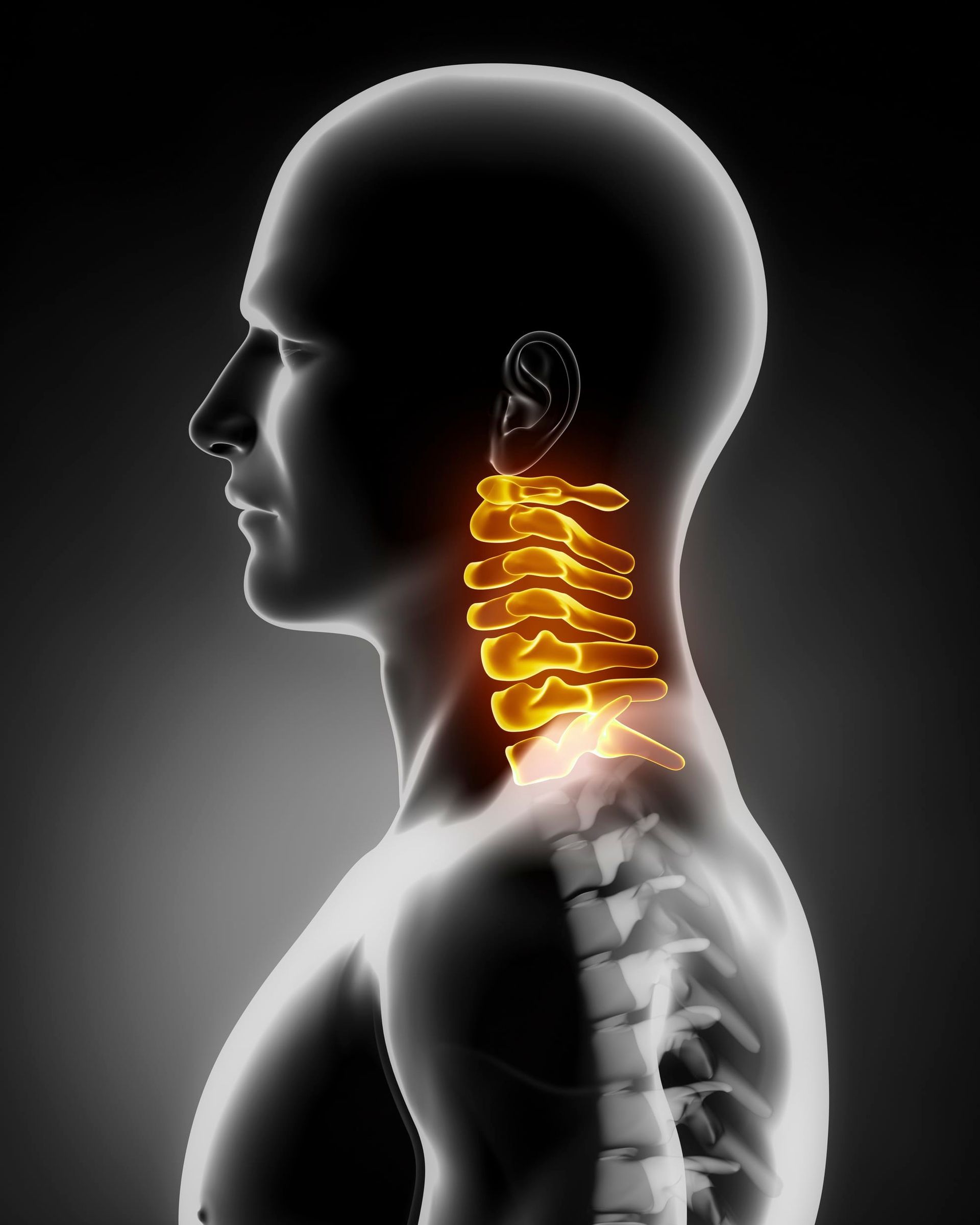
Cervical Conditions General Symptoms
Depending on the conditions, symptoms for Cervical Spine Conditions can range from low-grade pain of a stiff neck, numbness, tingling, or even weakness in the neck, arms, or shoulders as a result of pinched or irritated nerves in the cervical area.
Cervical Spine Disc Replacement
Cervical spine disc replacement surgery is a procedure that involves removing a degenerated or damaged cervical disc and replacing it with a device or artificial disc. Cervical discs provide cushion between the bones in your spine and neck (cervical spine) area. If a disc moves out of position, in even the slightest, it can cause pressure on the nerve root or on the spinal cord. This can cause neurological symptoms leading to pain, numbness and/or weakness.
Types of Cervical Conditions
A spinal tumor is an abnormal mass of tissue within or surrounding the spinal cord and spinal column, in which cells grow and multiply uncontrollably, seemingly unchecked by the mechanisms that control normal cells. Cervical Tumors can be benign (non-cancerous) or malignant (cancerous).
CAUSES
The cause of most primary spinal tumors is unknown. Some of them may be attributed to exposure to cancer-causing agents. Spinal cord lymphomas, which are cancers that affect lymphocytes, a type of immune cell, are more common in people with compromised immune systems. There appears to be a higher incidence of spinal tumors in particular families, so there is most likely a genetic component.
SYMPTOMS
Depending on the location and type of tumor, signs and symptoms include:
- Loss of sensation or muscle weakness, in the legs, arms or chest
- Difficulty walking, which may cause falls
- Decreased sensitivity to pain, heat and cold
- Loss of bowel or bladder function
- Paralysis that may occur in varying degrees and in different parts of the body, depending on which nerves are compressed
- Scoliosis or other spinal deformity resulting from a large but benign tumor
DIAGNOSIS
Initial treatment will involve thorough medical history and physical exam, including the patient’s symptoms. To help detect Cervical Tumors Robert Louis, MD conducts a neurological exam and may request for diagnostic imaging to find the exact location of disc degeneration causing the symptoms of Cervical Osteoarthritis.
Computerized tomography (CT Scan): CT scan may aid in determining the exact location of disc causing the nerve root and spinal cord compression.
X-ray: Application of radiation to produce a film or picture of a part of the body can show the structure of the vertebrae and the outline of the joints.
Magnetic resonance imaging (MRI): Advanced cases of Cervical Osteoarthritis show abnormal signal within the spinal cord on MRI imaging. In case of atrophy of the spinal cord due to nerve cell loss, also referred to as myelomalacia, surgical outcomes may not be as promising.
TREATMENT
Because most of these tumors arise from advanced cancer from another organ, the goal of spinal treatment is usually to:
- Control the severe pain that often occurs with these tumors (e.g. by removing pressure on the nerve roots)
- Preserve neurological function (e.g. by removing the pressure on the spinal cord)
- Fix structural instability in the spine (e.g. by reconstruction unstable spine with a spinal fusion)
Minimally Invasive Spine Surgery Robert Louis, MD specializes in minimally invasive spine surgery, wherein he incorporates his education, experience and training in cutting edge technology and instrumentation.
A herniated disc is a condition that can involve any part of the spine, but it most commonly occurs in the neck (cervical spine).
CAUSES
As part of our aging process, our bones, muscles, joints and soft tissues normally start to degenerate. As our spine degeneration begins on the spine, it can affect the discs that cushion and separate the vertebrae, leading to the formation of cracks and fissures in the disc walls.
SYMPTOMS
The symptoms of a herniated disc depend on the exact level of the spine where the disc herniation occurs and whether or not nerve tissue is being irritated. A disc herniation may not cause any symptoms. However, disc herniation can cause local pain at the level of the spine affected. Some of these symptoms include:
- Shooting pain down the buttock into the back of the thigh and down the leg
- Numbness and tingling in the leg.
- Pain often is worsened upon standing and decreases with lying down
DIAGNOSIS
Initial treatment will involve thorough medical history and physical exam, including the patient’s symptoms. To help detect a herniated disc, Robert Louis, MD conducts a neurological exam and may request for diagnostic imaging to find the exact location of disc degeneration causing the symptoms of a herniated disc.
Computerized tomography (CT Scan): CT scan may aid in determining the exact location of disc causing the nerve root and spinal cord compression.
Magnetic resonance imaging (MRI): Advanced cases of Cervical Osteoarthritis show abnormal signal within the spinal cord on MRI imaging. In case of atrophy of the spinal cord due to nerve cell loss, also referred to as myelomalacia, surgical outcomes may not be as promising.
X-ray: Application of radiation to produce a film or picture of a part of the body can show the structure of the vertebrae and the outline of the joints.
TREATMENT
Depending on the severity of symptoms, the treatments Dr. Louis would recommend for a herniated disc include physical therapy, muscle-relaxant medications, pain medications, anti-inflammation medications, local injection of cortisone (epidural injections), and surgical operations. Surgical options include microdiscectomy using small surgical instruments and open surgical repair.
Minimally Invasive Spine Surgery Robert Louis, MD specializes in minimally invasive spine surgery, wherein he incorporates his education, experience and training in cutting edge technology and instrumentation.
Causes of neck pain include:
CAUSES
- Abnormalities in the bone or joints
- Trauma
- Poor posture
- Degenerative diseases
- Tumors
- Muscle strain
SYMPTOMS
Symptoms that may occur with neck pain include neck tenderness, neck muscle spasm, and neck stiffness. Symptoms associated with serious causes of neck pain include arm weakness, leg weakness, arm numbness, leg numbness, loss of bladder control, loss of bowel control, and an inability to walk.
DIAGNOSIS
Initial treatment will involve thorough medical history and physical exam, including the patient’s symptoms. To help detect the source of the neck pain, Robert Louis, MD conducts a neurological exam and may request for diagnostic imaging to find the exact location of disc degeneration causing the symptoms of Neck Pain.
Computerized tomography (CT Scan): CT scan may aid in determining the exact location of disc causing the nerve root and spinal cord compression.
Magnetic resonance imaging (MRI): Advanced cases of Cervical Osteoarthritis show abnormal signal within the spinal cord on MRI imaging. In case of atrophy of the spinal cord due to nerve cell
loss, also referred to as myelomalacia, surgical outcomes may not be as promising.X-ray: Application of radiation to produce a film or picture of a part of the body can show the structure of the vertebrae and the outline of the joints.
Electrodiagnostic studies: Electromyography (EMG) and nerve conduction velocity (NCV) are sometime used to diagnose neck and shoulder pain, arm pain, numbness, and tingling.
TREATMENT
Initial treatments for neck pain are usually medications, surgery, therapy, and non-surgical, such as injections. Surgery may be needed for relieving nerve root or spinal cord compression.
Minimally Invasive Spine Surgery Robert Louis, MD specializes in minimally invasive spine surgery, wherein he incorporates his education, experience and training in cutting edge technology and instrumentation.
Radiculopathy refers to symptoms resulting from compression or irritation of the spinal nerve root at its point of connection to the spinal column. This injury can also be referred to as a pinched nerve.
CAUSES
Radiculopathy is caused by compression or irritation of the nerves as they exit the spine. This can be due to mechanical compression of the nerve by a disc herniation, a bone spur (osteophytes) from osteoarthritis, or from thickening of surrounding ligaments.
SYMPTOMS
The symptoms of radiculopathy depend on which nerves are affected. Symptoms of Radiculopathy include:
- Numbness and tingling in the arms or legs
- Localized neck or back pain
- Hypersensitivity to light touch that feels painful in the area involved
- Weakness and loss of coordination
DIAGNOSIS
Initial treatment will involve thorough medical history and physical exam, including the patient’s symptoms. To help detect Radiculopathy, Robert Louis, MD conducts a neurological exam and may request for diagnostic imaging to find the exact location that’s causing the symptoms of Radiculopahy such as an MRI which is the gold standard for providing the highest level of imaging of the spine, a CT Scan which may also be helpful in examining bone quality alignment and evaluation of fractures and other conditions, or an EMG which looks at the electrical activity along the nerve and can show if there is damage to the nerve.
TREATMENT
Most people can obtain good relief of their symptoms of radiculopathy with conservative treatment. This may include anti-inflammatory medications, physical therapy or chiropractic treatment, and avoiding activity that strains the neck or back. The majority of radiculopathy patients respond well to this conservative treatment, and symptoms often improve within 6 weeks to 3 months. If patients do not improve with the treatments listed above they may benefit from an epidural steroid injection. If treatments are unsuccessful and the symptoms are severe, surgery may be an option. The goal of the surgery is to remove the compression from the affected nerve. Depending on the cause of the radiculopathy, this can be done by a laminectomy or a discectomy.
Robert Louis, MD specializes in minimally invasive spine surgery, wherein he incorporates his education, experience and training in cutting edge technology and instrumentation. Below are the treatment options for radiculopathy:
- Anterior Cervical Discectomy and Fusion (ACDF)
- Cervical Disc Replacement
- Cervical Decompression
Spinal stenosis is the narrowing of the space for the spinal cord or nerve branches. More specifically, as the spine degenerates over time, it can lead to the formation of bone spurs.
CAUSES
Spinal stenosis is most common in those over the age of 50 year old due to changes that occur in the spine as a person ages. As people get older, the bands of tissues that support the spine get thick and hard, bones and joints get bigger, and surfaces of the bones may buldge out.
SYMPTOMS
Symptoms of spinal stenosis either don’t show or gradually appear and get worse over time. Signs of spinal stenosis include:
- Pain the the neck or back
- Numbness, weakness, cramping, or pain in the arms or legs
- Pain that triggers down the leg and foot problems
- Pressure on nerves in the lower back
DIAGNOSIS
Initial treatment will involve thorough medical history and physical exam, including the patient’s symptoms. To help detect spinal stenosis, Robert Louis, MD conducts a neurological exam and may request for diagnostic imaging to find the exact location of disc or joint degeneration causing the symptoms of spinal stenosis.
Computerized tomography (CT Scan): CT scan may aid in determining the exact location of disc causing the nerve root and spinal cord compression.
Magnetic resonance imaging (MRI): Advanced cases of Cervical Osteoarthritis show abnormal signal within the spinal cord on MRI imaging. In case of atrophy of the spinal cord due to nerve cell loss, also referred to as myelomalacia, surgical outcomes may not be as promising.
X-rays: Application of radiation to produce a film or picture of a part of the body can show the structure of the vertebrae and the outline of the joints.
Myelogram: A test in which the docor injects liquid dye into your spinal column.
Bone scan: A test in which you are given a shot of radioactive substance that shows where bone is breaking down or being formed.
TREATMENT
Initial treatments for spinal stenosis are usually medications, non-surgical, such as injections, and physical therapy. Surgery may be needed to restore functionality and alleviate pain in the affected joints or discs. If the pain worsens and treatments are unsucessful then surgery will be taken into consideration.
Minimally Invasive Spine Surgery Robert Louis, MD specializes in minimally invasive spine surgery, wherein he incorporates his education, experience and training in cutting edge technology and instrumentation.
Cervical myelopathy is a condition where the spinal cord in the neck (cervical spine) becomes compressed or damaged, usually due to degenerative changes in the spine.
CAUSES
The aging process results in degenerative changes in the cervical spine that, in advanced stages, can cause compression of the spinal cord resulting in cervical myelopathy. As you age, the bones and cartilage that make up your backbone and neck gradually develop wear and tear. These changes include dehydrated discs, herniated discs, bone spurs, and stiff ligaments.
SYMPTOMS
For most people cervical myelopathy causes no symptoms. When symptoms do occur, they typically include pain and stiffness in the neck. Sometimes, cervical spondylosis results in a narrowing of the space needed by the spinal cord and the nerve roots that pass through the spine to the rest of your body. Some of these symptoms include:
- Tingling, numbness and weakness in your arms, hands, legs or feet
- Lack of coordination and difficulty walking
- Stiffness and pain in the morning, then improves after getting up and moving around
- Loss of bladder or bowel control
DIAGNOSIS
Initial treatment will involve thorough medical history and physical exam, including the patient’s symptoms. To help detect cervical myelopathy, Robert Louis, MD conducts a neurological exam and may request for diagnostic imaging to find the exact location of nerve damage causing the symptoms of cervical myelopathy.
Computerized tomography (CT Scan): CT scan may aid in determining the exact location of disc causing the nerve root and spinal cord compression.
Magnetic resonance imaging (MRI): Advanced cases of Cervical Osteoarthritis show abnormal signal within the spinal cord on MRI imaging. In case of atrophy of the spinal cord due to nerve cell loss, also referred to as myelomalacia, surgical outcomes may not be as promising.
Myelogram: A test in which the docor injects liquid dye into your spinal column.
Somatosensory Evoked Potentials (SSEP): An electrical study done by stimulating the arms/legs and then reading the signal in the brain.
TREATMENT
The only effective cervical myelopathy treatment is surgical decompression of the spinal canal. Cervical stenosis surgical decompression can be performed through an anterior (front) approach or posterior (back) approach. The type of approach for cervical spinal stenosis is generally dependent on where Dr. Louis’s see’s the majority of the compression is located.
Minimally Invasive Spine Surgery Robert Louis, MD specializes in minimally invasive spine surgery, wherein he incorporates his education, experience and training in cutting edge technology and instrumentation.
Cervical spondylosis is a general condition that occurs with aging and results in the weakening and drying out of the intervertebral discs in the neck.
Suffering from Painful Cervical Spine Conditions or Neck Pain in Orange County?
Robert Louis, MD, a fellowship-trained Orange County Neurosurgeon, has particular expertise in endoscopic and minimally invasive treatment of Cervical Spine Conditions. For appointment, please call (949) 383-4185 or Contact Us.
CONTACT US
Please feel free to fill out the form provided on the right with your questions and Dr. Louis and his team will get back to you as soon as possible.