
Anterior Cervical Discectomy and Fusion
Robert Louis, MD specializes in minimally invasive of Brain Tumor Surgery in Orange County, California. Dr. Louis, a fellowship-trained Orange County Neurosurgeon, is the Director of the Skull Base and Pituitary Tumor Program at Hoag Memorial Hospital in Orange County, California. He has particular expertise in endoscopic and minimally invasive treatment of benign and malignant brain tumors, sellar and parasellar tumors and skull base tumors. Through the use of cutting-edge neuroimaging and neuro-navigational equipment, Dr. Louis utilizes the concept of keyhole neurosurgery, which minimizes the damage to vascular and soft tissues surrounding the brain.
Discectomy Procedure
Discectomy is the surgical removal of a bulging or degenerative disc. It is performed anywhere along the spine from the neck (cervical) to the low back (lumbar). Dr. Louis reaches the damaged disc from the front (anterior) of the spine through the throat area, moving aside the neck muscles, trachea, and esophagus to expose the disc and vertebrae. An anterior approach is used to reach the disc reached without disturbing the spinal cord, spinal nerves, and neck muscles. Depending on your symptoms and condition, one disc (single-level) or more (multi-level) may be removed.
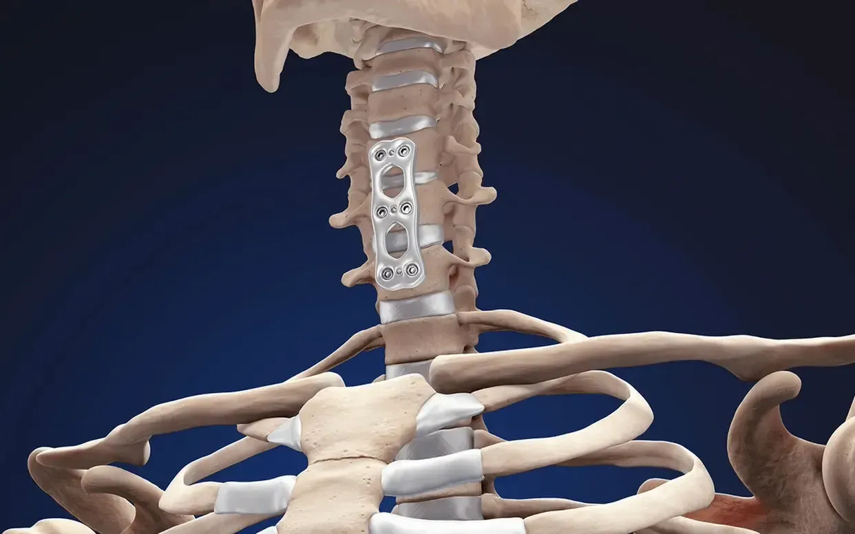
Fusion Procedure
The space between the vertebrae is empty after the affected disc is removed. To prevent the vertebrae from collapsing and rubbing together, a bone graft or bone graft substitute is inserted to create a spinal fusion and fill the open disc space. The bone graft and vertebrae are fixed in place with metal plates and screws for reinforcement. Following surgery, the body begins its natural healing process and new bone cells grow around the graft. After three to six months, the bone graft should join the two vertebrae and form one solid piece of bone. After fusion, the patient may notice some loss in the range of motion, which varies according to neck mobility before the fusion and the number of levels fused.
Types of Bone Graft
Autograft– The bone graph comes from the patient. Dr. Louis takes your own bone cells from the hip (iliac crest). This graft has a higher success rate of fusion because it has bone-growing cells and proteins. Harvesting a bone graft from your hip is done at the same time as spine surgery. The harvested bone is about a half-inch thick.
Allograft– The bone comes from a donor (cadaver) and is collected from people who have agreed to donate their organs after they die. This type of bone graft does not have bone-growing cells or proteins, yet it is readily available and eliminates the need to harvest bone from your hip. The center of the allograft is filled with shavings of living bone tissue taken from your spine during surgery.
Bone Graft Substitute– This type of graft is made of plastic, ceramic, or bioabsorbable substitute materials. Also called cages, bo graft material is filled with shavings of living bone tissue taken from your spine during surgery. For more information on ACDF, please call Dr. Louis at (949) 383-4185 or Contact Us.